X-ray tomography & Interferometry
Tomography is 3D imaging of objects from shadow cast by a beam. At CAMD(Center for the Advanced Microstructures and Devices), we use monochromatic X-rays, varies up to 60 keV to image with an effective spatial resolution of 3 µm at best. The field of view is determined by the X-ray beam height ( 1-3 mm ) and the pixel number in the sCMOS camera (2k x 2k). So, CAMD can image a volume of 3 mm x 5 mm x 5 mm at 3 µm resolution. Because of the math of tomography, the object should have a diameter smaller than 5 mm. The speed of tomography imaging ranges for1 to 2 hrs, depending upon camera exposure time (up to10 seconds) and number of projections used to create the 3D image. The samples must be translucent to the X-rays; this can force samples to be even smaller than 5 mm. The image contrast is a function of variations in X-ray absorption through the sample: air-material structure is easy; tissue-tissue interfaces will be required added contrast agents.
Beamline is also equipped with three gratings to perform Talbot Lau interferometry imaging. Currently we are conducting x-ray absorption tomography and 2D/3D- Interferometry imaging with X-ray Energy > 35keV.
Beamline also offers new instrument feasibility test, such as innovated grating system test and in-situ sample environmental change observation test(heat).
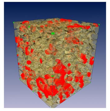 Porous Media
Kyungmin Ham at 225-578-9350 or kham1@lsu.edu